
Heel Pain & Heel Fractures
Plantar Fasciitis and Heel Pain
Heel pain is most often caused by plantar fasciitis, a condition that is sometimes also called heel spur syndrome when a spur is present. Heel pain may also be due to other causes, such as a stress fracture, tendonitis, arthritis, nerve irritation, or, rarely, a cyst. Because there are several potential causes, it is important to have heel pain properly diagnosed. A foot and ankle surgeon is able to distinguish between all the possibilities and determine the underlying source of your heel pain.

What Is Plantar Fasciitis?
Plantar fasciitis is an inflammation of the band of tissue (the plantar fascia) that extends from the heel to the toes. In this condition, the fascia first becomes irritated and then inflamed, resulting in heel pain.
Causes
The most common cause of plantar fasciitis relates to faulty structure of the foot. For example, people who have problems with their arches, either overly flat feet or high-arched feet, are more prone to developing plantar fasciitis.
Wearing non-supportive footwear on hard, flat surfaces puts abnormal strain on the plantar fascia and can also lead to plantar fasciitis. This is particularly evident when one’s job requires long hours on the feet. Obesity may also contribute to plantar fasciitis.
Symptoms
The symptoms of plantar fasciitis are:
- Pain on the bottom of the heel
- Pain that is usually worse upon arising
- Pain that increases over a period of months
People with plantar fasciitis often describe the pain as worse when they get up in the morning or after they’ve been sitting for long periods of time. After a few minutes of walking the pain decreases, because walking stretches the fascia. For some people the pain subsides but returns after spending long periods of time on their feet.
Diagnosis
To arrive at a diagnosis, the foot and ankle surgeon will obtain your medical history and examine your foot. Throughout this process the surgeon rules out all the possible causes for your heel pain other than plantar fasciitis.
In addition, diagnostic imaging studies such as x-rays or other imaging modalities may be used to distinguish the different types of heel pain. Sometimes heel spurs are found in patients with plantar fasciitis, but these are rarely a source of pain. When they are present, the condition may be diagnosed as plantar fasciitis/heel spur syndrome.
Non-Surgical Treatment
Treatment of plantar fasciitis begins with first-line strategies, which you can begin at home:
- Stretching exercises.Exercises that stretch out the calf muscles help ease pain and assist with recovery.
- Avoid going barefoot.When you walk without shoes, you put undue strain and stress on your plantar fascia.
- Ice.Putting an ice pack on your heel for 20 minutes several times a day helps reduce inflammation. Place a thin towel between the ice and your heel; do not apply ice directly to skin.
- Limit activities. Cut down on extended physical activities to give your heel a rest.
- Shoe modifications. Wearing supportive shoes that have good arch support and a slightly raised heel reduces stress on the plantar fascia.
- Medications.Oral nonsteroidal anti-inflammatory drugs (NSAIDs), such as ibuprofen, may be recommended to reduce pain and inflammation.
If you still have pain after several weeks, see your foot and ankle surgeon, who may add one or more of these treatment approaches:
- Padding and strapping. Placing pads in the shoe softens the impact of walking. Strapping helps support the foot and reduce strain on the fascia.
- Orthotic devices. Custom orthotic devices that fit into your shoe help correct the underlying structural abnormalities causing the plantar fasciitis.
- Injection therapy. In some cases, corticosteroid injections are used to help reduce the inflammation and relieve pain.
- Removable walking cast. A removable walking cast may be used to keep your foot immobile for a few weeks to allow it to rest and heal.
- Night splint. Wearing a night splint allows you to maintain an extended stretch of the plantar fascia while sleeping. This may help reduce the morning pain experienced by some patients.
- Physical therapy. Exercises and other physical therapy measures may be used to help provide relief.
When Is Surgery Needed?
Although most patients with plantar fasciitis respond to non- surgical treatment, a small percentage of patients may require surgery. If, after several months of non-surgical treatment, you continue to have heel pain, surgery will be considered. Your foot and ankle surgeon will discuss the surgical options with you and determine which approach would be most beneficial for you.
Long-term Care
No matter what kind of treatment you undergo for plantar fasciitis, the underlying causes that led to this condition may remain. Therefore, you will need to continue with preventive measures. Wearing supportive shoes, stretching, and using custom orthotic devices are the mainstay of long-term treatment for plantar fasciitis.
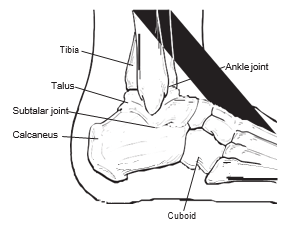
What is the Calcaneus?The calcaneus, also called the heel bone, is a large bone that forms the foundation of the rear part of the foot. The calcaneus connects with the talus and cuboid bones. The connection between the talus and calcaneus forms the subtalar joint. This joint is important for normal foot function.
The calcaneus is often compared to a hard boiled egg, because it has a thin, hard shell on the outside and softer, spongy bone on the inside. When the outer shell is broken, the bone tends to collapse and become fragmented. For this reason, calcaneal fractures are severe injuries. Furthermore, if the fracture involves the joints, there is the potential for long-term consequences such as arthritis and chronic pain.
How do Calcaneal Fractures Occur?
Most calcaneal fractures are the result of a traumatic event— most commonly, falling from a height, such as a ladder, or being in an automobile accident where the heel is crushed against the floorboard. Calcaneal fractures can also occur with other types of injuries, such as an ankle sprain. A smaller number of calcaneal fractures are stress fractures, caused by overuse or repetitive stress on the heel bone.
Types of Calcaneal Fractures
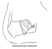
Fractures of the calcaneus may or may not involve the subtalar and surrounding joints. Fractures involving the joints (intra-articular fractures) are the most severe calcaneal fractures, and include damage to the cartilage (the connective tissue between two bones). The outlook for recovery depends on how severely the calcaneus was crushed at the time of injury.
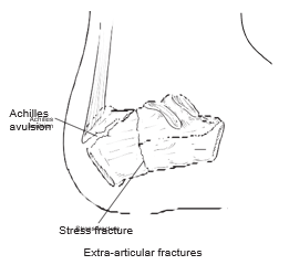
Fractures that don’t involve the joint (extra-articular fractures) include:
- Those caused by trauma, such as avulsion fractures (in which a piece of bone is pulled off of the calcaneus by the Achilles tendon or a ligament) or crush injuries resulting in multiple fracture fragments.
- Stress fractures, caused by overuse or mild injury.
The severity and treatment of extra-articular fractures depend on their location and size.
Signs and Symptoms
Calcaneal fractures produce different signs and symptoms, depending on whether they are traumatic or stress fractures.
The signs and symptoms of traumatic fractures may include:
- Sudden pain in the heel and inability to bear weight on that foot
- Swelling in the heel area
- Bruising of the heel and ankle
The signs and symptoms of stress fractures may include:
- Generalized pain in the heel area that usually develops slowly (over several days to weeks)
- Swelling in the heel area
Diagnosis
To diagnose and evaluate a calcaneal fracture, the foot and ankle surgeon will ask questions about how the injury occurred, examine the affected foot and ankle, and order x-rays. In addition, advanced imaging tests are commonly required.
Treatment
Treatment of calcaneal fractures is dictated by the type of fracture and extent of the injury. The foot and ankle surgeon will discuss with the patient the best treatment— whether surgical or non-surgical— for the fracture.
For some fractures, non-surgical treatments may be used. These include:
- Rest, ice, compression, and elevation (R.I.C.E.) Rest (staying off the injured foot) is needed to allow the fracture to heal. Ice reduces swelling and pain; apply a bag of ice covered with a thin towel to the affected area. Compression (wrapping the foot in an elastic bandage or wearing a compression stocking) and elevation (keeping the foot even with or slightly above the heart level) also reduce the swelling.
- Immobilization. Sometimes the foot is placed in a cast or cast boot to keep the fractured bone from moving. Crutches may be needed to avoid weightbearing.
For traumatic fractures, treatment often involves surgery to reconstruct the joint, or in severe cases, to fuse the joint. The surgeon will choose the best surgical approach for the patient.
Rehabilitation
Whether the treatment for a calcaneal fracture has been surgical or non-surgical, physical therapy often plays a key role in regaining strength and restoring function.
Complications of Calcaneal Fractures
Calcaneal fractures can be serious injuries that may produce lifelong problems. Arthritis, stiffness, and pain in the joint frequently develop. Sometimes the fractured bone fails to heal in the proper position. Other possible long-term consequences of calcaneal fractures are decreased ankle motion and walking with a limp due to collapse of the heel bone and loss of length in the leg. Patients often require additional surgery and/or long term or permanent use of a brace or an orthotic device (arch support) to help manage these complications.